")
")
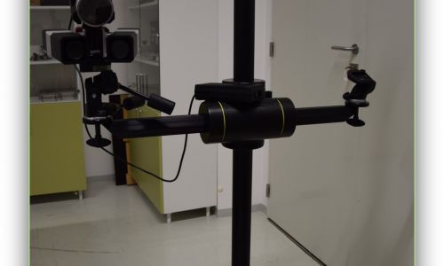
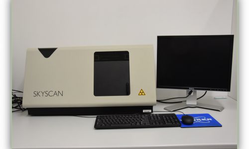
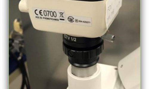
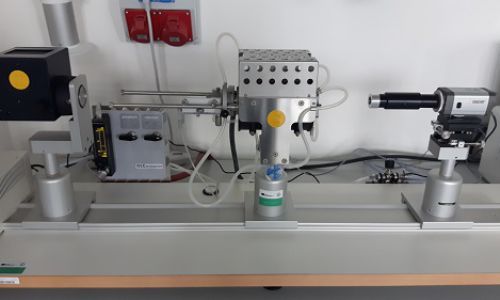
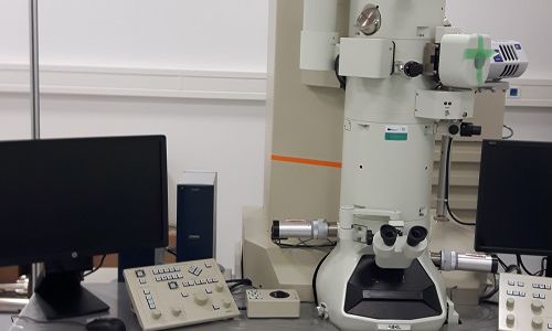
X-ray Tomograph 3D
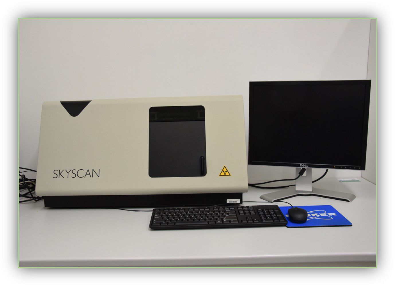
Device is for:
- Tomograph used to create a 3D image of the sample to be used to find defects in the sample (dirt, heteromaterial, air bubbles).
- The device operates on the basis of the density difference, which should be taken into account when using this technique.
- The machine's maximum resolution is 6-7 micrometers.
contact person:
doc. Ing. et doc. Ing. Ivo Kuřitka, Ph.D. et Ph.D.
+420 57 603 8049 A418
This email address is being protected from spambots. You need JavaScript enabled to view it.
research